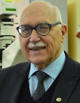
Prof. Valery Tuchin
Saratov State University, Russia
Speech Title: Advanced Biomedical
Imaging and Tomography at Tissue Optical Clearing
Abstract: Principles and novelties in
the field of tissue optics in context of tissue optical
clearing (TOC) will be presented. New fields of clinical
applications for optical imaging, monitoring of drug
delivery, effective antitumor and antimicrobial
phototherapy, optical communication with implants in the
human body, and laser therapy and nanosurgery will be
discussed.
TOC is based on temporal suppression of tissue light
scattering using immersion optical clearing agents
(OCAs) [1-4], which make the living tissue to be
reversibly transparent in a wide spectral range from
deep UV to terahertz, providing a higher imaging depth
and a better contrast in optics, CT and MRI.
1. L. Oliveira and V. V. Tuchin, The Optical Clearing
Method: A New Tool for Clinical Practice and Biomedical
Engineering, Springer Nature Switzerland AG, Basel, 2019
– 177 p.
2. V. V. Tuchin, D. Zhu, and E. A. Genina (Eds.),
Handbook of Tissue Optical Clearing: New Prospects in
Optical Imaging, Taylor & Francis Group LLC, CRC Press,
Boca Raton, FL, 2022 – 688 p.
3. V.V. Tuchin, E.A. Genina, E.S. Tuchina, A.V.
Svetlakova, Y.I. Svenskaya, Optical clearing of tissues:
issues of antimicrobial phototherapy and drug delivery,
Advanced Drug Delivery Reviews 180 (1), 114037 (2022).
4. I.S. Martins, H.F. Silva, E.N. Lazareva, N.V.
Chernomyrdin, K.I. Zaytsev, L.M. Oliveira, and V.V.
Tuchin, Measurement of tissue optical properties in a
wide spectral range: a review [Invited], Biomedical
Optics Express, 14 (1), 249-298 (2023).
Bio-sketch: Valery V. Tuchin is the
corresponding member of the Russian Academy of Sciences,
Head of the Department of Optics and Biophotonics and
Director of the Science Medical Center of Saratov State
University. He is also works with several other research
centers of RAS and universities. His research interests
include biophotonics, biomedical optics, and
nanobiophotonics. He is a Honored Scientist of Russia,
Fellow of SPIE and OPTICA, Distinguished Professor of
Finland, recipient of SPIE Educator Award, Joseph W.
Goodman Book Writing Award (OPTICA/SPIE), Michael S.
Feld Biophotonics Award (OPTICA), Chime Bell Award of
the Chinese province of Hubei, the D.S. Rozhdestvensky
Medal and the S.I. Vavilov Medal of the Russian Optical
Society, and the A.M. Prokhorov Medal of the Russian
Academy of Engineering Sciences.

Prof. Ehrenfried Zschech
DeepXscan GmbH, Germany
Speech Title: High-resolution X-ray
Imaging – Challenges to Experiment and Data Analysis
Abstract: High-resolution X-ray imaging
provides nondestructive characterization capabilities on
opaque objects, observing features with sizes across a
range of length scales, down to several 10 nanometers
using lens-based transmission X-ray microscopy (TXM).
Multi-scale X-ray computed tomography (XCT),
characterized by a sample thickness / resolution value
of ~ 103, and subsequent 3D data reconstruction is an
efficient approach to study the 3D morphology of natural
and engineered hierarchically structured systems and
materials. Because of the ability of micro-XCT and
nano-XCT to reveal structural characteristics and
materials’ microstructure, they are potential imaging
techniques for micro- und nano-structured objects
(microchips), advanced multi-component materials
(composites and skeleton materials) as well as
biological objects (diatoms or mollusk shells). In this
talk, multi-scale imaging of the 3D morphology of a
novel, hierarchically structured transition-metal-based
electrocatalytic system for water splitting, applying
laboratory high-resolution multi-scale XCT, will be
demonstrated.
Lens-based TXM and nano-XCT have been demonstrated at
synchrotron radiation (SR) beamlines but also in the
laboratory, with 24/7 tool access. Since the photon flux
of laboratory X-ray sources is several orders of
magnitude less compared to synchrotrons, image analysis
including denoising is needed to get high-quality 3D
information of the object. While XCT is pushed further
into the micro- and nanoscale, the mitigation of
artefacts caused by thermomechanical instability of tool
components and object motion, by center of rotation
misalignment and by inaccuracy in the detector position
require computational efforts, e.g. the application of
deep convolutional neural networks. Therefore, 3D
reconstruction methodologies are needed that consider
unavoidable misalignment and object motion during the
data acquisition, in order to obtain high-quality 3D
images [1,2].
[1] E. Topal et al., BMC Mater. 2, 1 (2020), Sci. Rep.
10, 1 (2020)
[2] K. Bulatov et al., Nanomaterials 11, 2524 (2021)
Bio-sketch: Ehrenfried Zschech is CTO and Co-Founder of deepXscan GmbH, Dresden, Germany. His responsibilities include research and innovation in the field of high-resolution X-ray imaging and the development of customized solutions for a broad range of applications including advanced packaging for microelectronics. He is member of the European Academy of Science (EurASc) and of ACATECH Germany. Ehrenfried Zschech had several management positions at Airbus in Bremen, at Advanced Micro Devices in Dresden and at Fraunhofer. He holds honorary professorships for Nanomaterials at Brandenburg University of Technology Cottbus-Senftenberg and for Nanoanalysis at Dresden University of Technology. Ehrenfried Zschech was awarded with the FEMS European Materials Gold Medal in 2019.

Prof. Victor A. Soifer
Samara University, Russia
Bio-sketch: Academician of RAS Victor
Soifer, Dr.Sc. (1979), PhD (1971), President of Samara
National Research University, the chief researcher of
the IPSI RAS –Branch of the FSRC “Crystallography and
Photonics” RAS and the Editor-in-Chief of Computer
Optics. He is a SPIE-and IAPR-member. He is the author
and coauthor of more than 700 scientific publications,
10 books, and 50 author’s certificates and patents.
General research areas: diffractive optics and
nanophotonics, image analysis, pattern recognition,
computer vision, and artificial intelligence.
Web of Science:
https://www.webofscience.com/wos/author/record/C-3088-2017
Scopus ID:
https://www.scopus.com/authid/detail.uri?authorId=36836834300
ORCID:
https://orcid.org/0000-0003-4239-4389
Invite Speaker

Dr. Flavio Piccoli
University of Milano, Italy
Speech Title: From Black-Box to
White-Box Image Enhancement: Leveraging Deep Learning
and User-Centric Adaptation
Abstract:
Image enhancement is a
critical field that spans machine vision and human-user
applications. This presentation centers on the concept
of black-box image enhancement, driven by deep learning
techniques, and the transformative journey towards
white-box solutions and user-centric adaptation.
Black-Box Image Enhancement: Explore the traditional
approach of black-box image enhancement, where
sophisticated algorithms often operate as opaque
processes, making it challenging to understand,
customize, or personalize their outputs. This
introductory section will set the stage for the
evolution to more transparent and user-centric
approaches.
White-Box Image Enhancement: Delve into the power of
interpretable approaches, where the output can be
further adapted to the needs of the user. Understand how
these advanced methods can be harnessed to bring
transparency and interpretability to the previously
black-box enhancement techniques.
User-Centric Adaptation: Uncover strategies for adapting
image enhancement algorithms to user preferences and
flavors. Learn how the combination of deep learning and
user-centric adaptation can transition image enhancement
from a black-box and white-box processes into
personalized algorithms, offering users the ability to
customize and personalize image outputs, ultimately
enhancing the visual experience for both machines and
humans.
Bio-sketch: Flavio
Piccoli is an Assistant Professor of Computer Science in
the Department of Excellence in Informatics, Systems and
Communication at the University of Milano-Bicocca. He is
also an associate member of the Imaging and Vision Lab
(IVL), directed by Prof. Raimondo Schettini.
From 2019 to 2023 he was a postdoctoral research fellow
at the Italian National Institute of Nuclear Physics
(INFN) and the University of Milano-Bicocca. In 2019, he
received the degree of Doctor of Philosophy degree (PhD)
in Computer Science with a thesis focused on automatic
quality inspection. In 2014, he received his master's
degree in Computer Science with a thesis focused on face
analysis methods. He spent a period abroad at the
Computer Vision Center (CVC) laboratory of the
Universitat Autònoma de Barcelona (UAB), in the Learning
and Machine Perception (LAMP) research group directed by
Prof. Joost van de Weijer.
His main research interests are in the field of
artificial intelligence, machine and deep learning,
computer vision, image enhancement, remote sensing, and
automatic quality inspection.
He has authored several scientific papers in indexed and
high Scopus-scoring journals and conferences. He is an
associate editor of the SPIE Journal of Electronic
Imaging and Frontiers in Artificial Intelligence. He is
currently teaching the course "Advanced Computational
Techniques for Big Imaging and Signal Data" for the
innovative inter-university master's degree course in
Artificial Intelligence for Science and Technology,
jointly organized by the University of Milan, the
University of Pavia and the University of
Milano-Bicocca.
Tutorial

Prof. Wolfgang Osten
Universität Stuttgart, German
Speech Title: On the Meaning and
Partial Misuse of Some frequently Applied Terms in
Optical Sensor Technologies
Abstract: Optical sensor technologies
both coherent and no-coherent are gaining continuously
more importance in all industrial and public branches.
That is a consequence of their unique features such as
the non-contact and high speed interaction with the
object under inspection, the largely free scalability of
the dimension of the probing tool, the high resolution
of the data, and the diversity of information channels
of the light field. With the progress of production
technologies and the innovation of new products with
outstanding features the challenges to all kind of
inspection devices are dramatically increasing. In this
tutorial we turn our attention tot he discussion of
terms that are currently used very common but that are
causing an expectation that is not (yet) really
realistic. To them belong such frequently used terms
such as „artificial intelligence“, „autonomous
vehicles“, „superresolution“, „digitization“ and
„holography“. We try to make clear what is meant when
using these terms, we show the potential but also the
challenges on example of selected applications. Overall,
this paper is not a scolding against the partly
inflationary use of these terms. On the contrary, it is
an attempt to objectify the discussion in order to avoid
frustration when the excessive expectations fail again.
Bio-sketch: Wolfgang Osten received the MSc/Diploma in Physics from the Friedrich-Schiller-University Jena in 1979. From 1979 to 1984 he was a member of the Institute of Mechanics in Berlin working in the field of experimental stress analysis and optical metrology. In 1983 he received the PhD degree from the Martin-Luther-University Halle-Wittenberg for his thesis in the field of holographic interferometry. From 1984 to 1991 he was employed at the Central Institute of Cybernetics and Information Processes ZKI in Berlin making investigations in digital image processing and computer vision. Between 1988 and 1991 he was heading the Institute for Digital Image Processing at the ZKI. In 1991 he joined the Bremen Institute of Applied Beam Technology (BIAS) to establish and to direct the Department Optical 3D-Metrology till 2002. Since September 2002 he has been a full professor at the University of Stuttgart and director of the Institute for Applied Optics. From 2006 till 2010 he was the vice rector for research and technology transfer of the Stuttgart University where he is currently the vice chair of the university council. His research work is focused on new concepts for industrial inspection and metrology by combining modern principles of optical metrology, sensor technology and image processing. Special attention is directed to the development of resolution enhanced technologies for the investigation of micro and nano structures.
